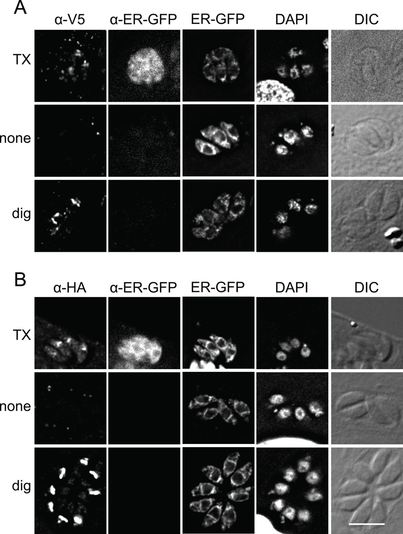
Figure 7. The N-terminus of APT1 faces the cytosol during trafficking.
T. gondii expressing V5t-APT1(Y16A)-HA and an ER-luminal GFP marker were fixed then permeabilized with Triton X-100, digitonin, or no detergent as indicated. Samples were then probed with anti-V5 mAb (A) or anti-HA mAb (B) as well as rabbit anti-GFP antibodies. The antibodies were premixed with Fab anti-mouse Ig coupled to Alexa 568 and Fab anti-rabbit Ig coupled to Alexa 680 respectively. Intrinsic fluorescence of ER-localized GFP and DAPI staining are also shown, along with DIC images. To facilitate proper scaling of the fluorescence signal, wild type parasites were included on each coverslips as a negative control (not shown). In some DAPI images, part of the host nucleus is visible. Bar = 5 µM.

