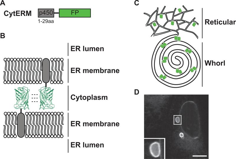
Figure 1. Expression of CytERM restructures the ER into OSER through oligomeric interactions of fluorescent proteins.
(A) CytERM, fusion illustration of amino acids 1-29 of cytochrome p450 with fluorescent protein. (B) Model of opposing ER membrane remodeling due to fluorescent protein oligomerization. (C) Illustration depicting reorganization of ER due to fluorescent protein interaction from typical reticular network to OSER whorl. (D) OSER whorl in U2OS cells transiently expressing CytERM-EGFP. Inset of OSER whorl. Scale bar = 10 μm.

