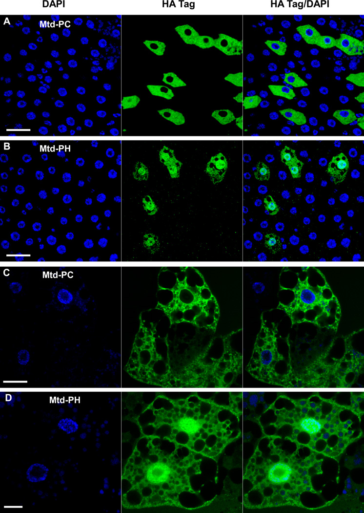
Figure 9. Mtd-PH is nuclearly localized, while Mtd-PC is excluded from the nucleus.
Immunofluorescence of Mtd-PC-HA (A) and Mtd-PH-HA (B) in the adult midgut. Immunofluorescence of Mtd-PC-HA (C) and Mtd-PH-HA (D) in the larval fat body. In all cases, expression was driven by Da-Gal4. The tagged protein is visualized with Alexa 488 (green), and nuclear DNA is stained with DAPI (blue). The mosaic pattern of expression is a characteristic of the Da-Gal4 driver. Scale bar = 20 µm.

