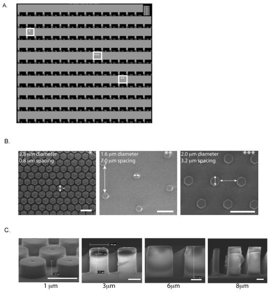
Figure 1. SiO2 micropillar substrates.
(A) Light microscope image of representative SiO2 micropillar substrate. SiO2 micropillar substrates were fabricated using I-line lithography followed by a timed reactive ion etch to create a unique substrate design presenting micropillar diameters ranging from 1-5.6 μm, with spacing between pillars ranging from 0.6-15 μm. (B) SEM images of various locations of the SiO2 micropillar substrate (indicated by stars), demonstrating wide array of design. (C) SEM images of SiO2 substrate of 1 μm, 3 μm, 6 μm, and 8 μm micropillar heights. Scale bar is 1 mm in (A), 5 μm in (B), and 2 μm in (C).

