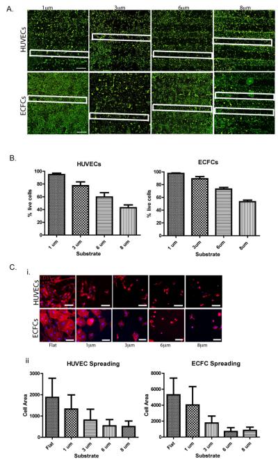
Figure 2. EC adhesion onto Fn-coated micropillared SiO2 substrates.
(A) HUVEC (upper panel) and ECFC (lower panel) adhesion onto Fn-coated micropillar SiO2 substrates with topographical feature heights ranging from 1-8 μm, cells stained with calcein (green-live staining). For clarification, some micropillar regions are outlined with a white box. (B) Quantification of percent of HUVEC (left) and ECFC (right) live cells on micropillar regions for each of the feature heights shows marked decreased percentage of live cells on substrates with topographical features with >3 μm heights. (C) HUVEC (upper panel) and ECFC (lower panel) expressing endothelial surface marker CD31 (red) have decreased cell spreading on substrates with topographical feature heights >1 μm as demonstrated by fluorescence microscope imaging (i) and cell spreading quantification (ii). Values shown are means ± SD. Scale bar is 1 mm in (A) and 100 μm in (C). Image analyses were performed on triplicate samples (n = 3).

