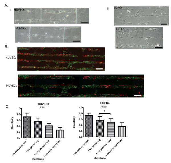
Figure 6. EC elongation of PDMS micropillar substrates.
(A) Light microscope images of HUVECs (i) and ECFCs(ii) demonstrate aligned and elongated morphologies after 24 h. (B) Flourescence microscope imaging of HUVECs stained with phalloidin (green) demonstrate successful patterning of PDMS micropillar substrates (Fn=red) and the preferential adhesion of HUVECs to Fn patterned regions. (C) Quantification of cell shape indicates significant HUVEC and ECFC elongation (a measure of circularity) on Fn-patterned SiO2 and PDMS micropillars compared to cells cultured on flat substrates. Significance levels were set at: *p < 0.05, **p < 0.01, and ***p < 0.001. Values shown are means ± SD.

