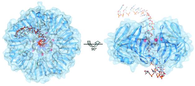
Figure 5.
Model of a SmAP⋅ssRNA complex. Orthogonal views are shown for a model of SmAP bound to a hypothetical 20-nucleotide ssRNA (e.g., eukaryotic snRNA) that consists of three segments from 5′ to 3′: a random string of 6 nucleotides in the A-form conformation (ACGAUC), followed by a minimal consensus Sm binding site (GAU4GA), and ending with 6 more nucleotides in A-form geometry (ACGAUC). The SmAP heptamer is depicted as a ribbon diagram along with the solvent-accessible surface. The Arg-29 ring that forms the pore is colored by atom type and rendered in space-filling form, with the ssRNA drawn as a stick model. The steric and electrostatic environment of the pore is ideally suited to accommodate a single-stranded polypyrimidyl nucleic acid.

