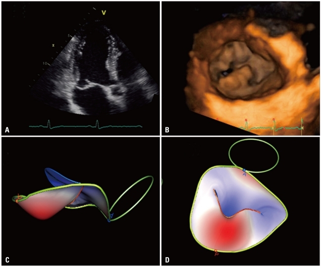
Fig. 5.
Degenerative mitral valve disease: A: Apical long-axis view showing a flail of posterior mitral leaflet. B: Volume rendering of the showing the location and extent of the prolapsing segment. C and D: Surface rendering of the valve leaflets, annulus and aortic annulus to provide an automated quantitative analysis of valve morphology and geometry useful to plan the surgical approach.

