
Figure 5.
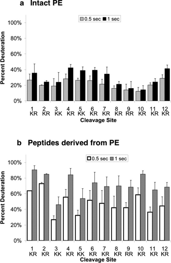
Differential H-D exchange among protease cleavage sites of PE and peptides derived from PE. (a) Intact PE: H-D exchange at cleavage site subdomains. Comparison of relative H-D exchange rates at the 12 dibasic residue cleavage sites of PE are illustrated at 0.5 and 1 s time points for PE incubation in D2O (hatched and black bars, respectively). H-D exchange data was compiled from multiple peptides of PE that span each of the cleavage sites (indicated in figure) and their average level of percent deuteration was calculated (mean ± s.e.m.). (b) Peptides derived from PE: H-D exchange of peptides spanning cleavage site domains. H-D exchange of peptides spanning multiple cleavage sites are illustrated for 0.5 and 1 s exchange time periods (white and gray, respectively). H-D exchange data for peptides spanning each of the cleavage sites (indicated in figure) were compiled and their average level of percent deuteration was calculated (mean ± s.e.m.). The standard deviation of deuterium incorporation measured in replicate determinations was typically less than 5% of the mean, as reported (49, 50).