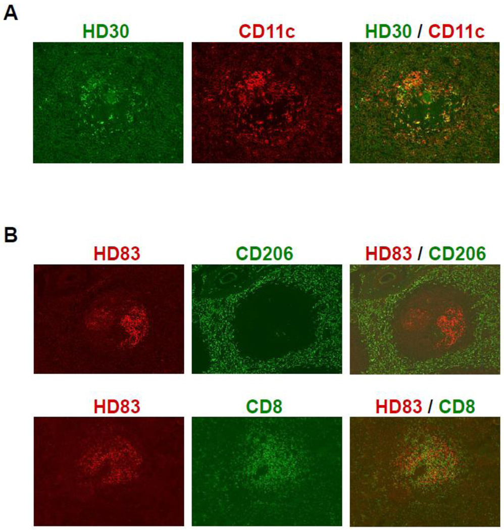
Figure 3. Immunohistochemical stains of HD mAbs on the splenic tissues from human and monkey.
(A) Cells expressing DEC205 at high levels were detected in the white pulp of the human spleen by HD30. Double staining of human spleen sections was carried out with anti-DEC205 mAb HD30 (in green) and anti-CD11c antibody (DCs in red).
(B) HD83 stained DEC205+ cells in the T-cell areas of the white pulps in monkey spleen. Double staining of monkey spleen sections was carried out with anti-DEC205 mAb HD83 (in red) and antibodies to CD206 (red pulp macrophages in green; upper panel) or CD8 (T cells in green; lower panel).

