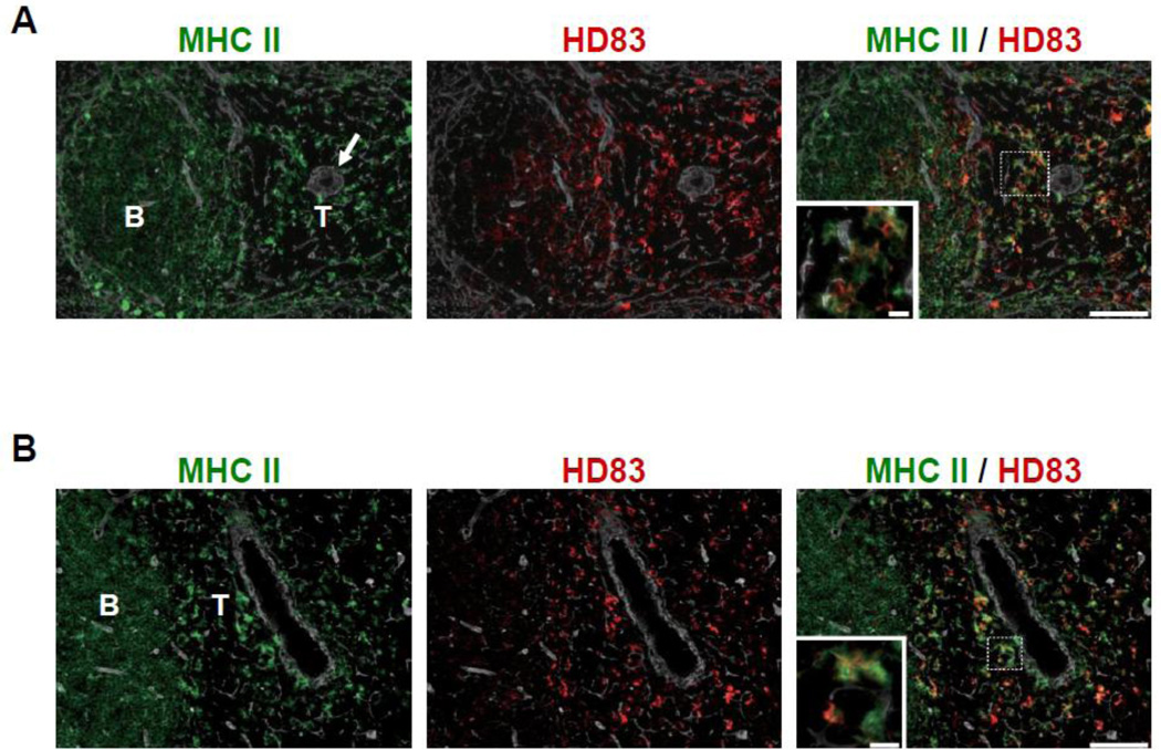
Figure 4. HD83 stained DEC205+ DCs in the T-cell areas of rat lymphoid tissues.
(A) Rat spleen was stained triple-immunofluorescently for HD83 (red), class II MHC (green), and tissue framework (IV collagen, white). Inset shows dendritic shape of HD83+ class II MHC+ cells in high magnification. Arrow indicates central artery. B: B-cell area; T: T-cell area; Scale bar = 80 µm (10 µm in high magnification).
(B) Rat cervical lymph node was stained as in (A).

