Figure 3.
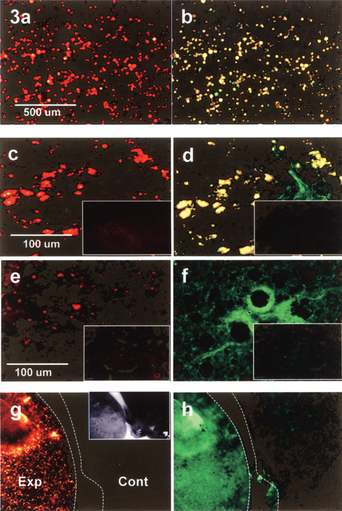
Sertoli cells (SCs) preloaded with FITC-labeled nanoparticles and prelabeled with DiI viewed through the TRITC filter (a, c, e, g) and through the FITC filter (b, d, f, h). Intact cells in vitro image red through the TRITC filter (a) and yellow through the FITC filter (b). Intact SCs in lung cryosections 15 min postinjection (c, d) image identically to the cells in vitro. However, at 1 h postinjection (e, f) note the absence of intact SCs (yellow) when viewed through the FITC filter but positive fluorescence for FITC-labeled nanoparticles (green). Cryosections from uninjected control mice (insets) show no positive fluorescence for SCs or nanoparticles. Low magnification macroscopic images of whole lung samples from mice 1 h postinjection (g, h Exp) imaged similarly to the sectioned lungs 1 h postinjection (e, f). SC label was present when viewed through the TRITC filter (g, Exp), but the absence of overlap fluorescence (yellow), when viewed through the FITC filter (h, Exp), indicated the absence of intact SCs but instead the presence of labeled nanoparticles (green). Whole lung samples from control mice (g, h Cont) showed no positive fluorescence. Inset in (g) shows the two whole lung samples (Exp and Cont) imaged by bright field stereoscopic microscopy.

