Figure 1.
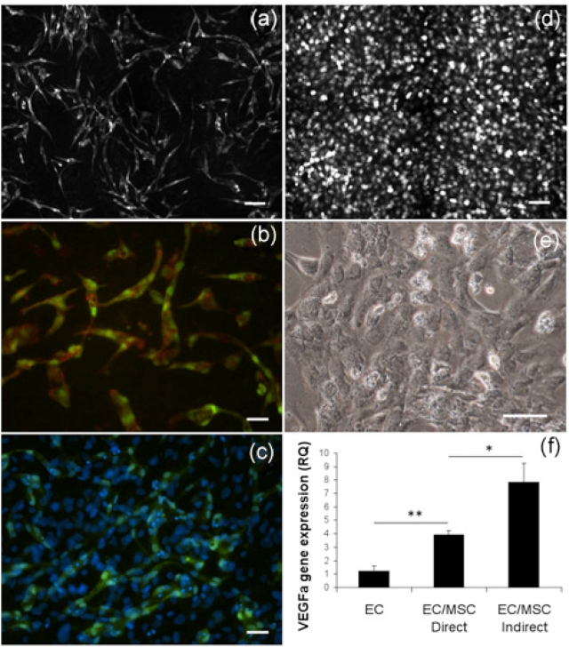
Endothelial network establishment in coculture with bone marrow–derived MSCs. (a) The 2 × 2 montage image (10×) with live fluorescence microscopy from network of GFP-expressing EC cocultured with MSC. At 6 days, ECs have organized into stabilized networks. Scale bar = 100 µm. (b) UEA lectin staining of fixed GFP-expressing EC (20×) organized in elongated structures at 6 days. Scale bar = 50 µm. (c) DAPI staining (20×) showing the confluent layer of MSC supporting EC network formation at 6 days. Scale bar = 50 µm. (d) Live fluorescence microscopy after 6 days of monocultured GFP-expressing EC (10×) that did not spontaneously organize into capillary-like networks. Scale bar = 100 µm. (e) Monocultured EC (20×) presented a cobblestone-like morphology. Scale bar = 50 µm. (f) EC expression of VEGFa was significantly upregulated in coculture at 3 days, but also significantly lower than in indirect culture with shared culture medium. *p < 0.05, **p < 0.01.
GFP: green fluorescent protein; EC: endothelial cell; MSC: mesenchymal stem cell; DAPI: 4′,6-diamidino-2-phenylindole; VEGF: vascular endothelial growth factor.

