
Figure 5.
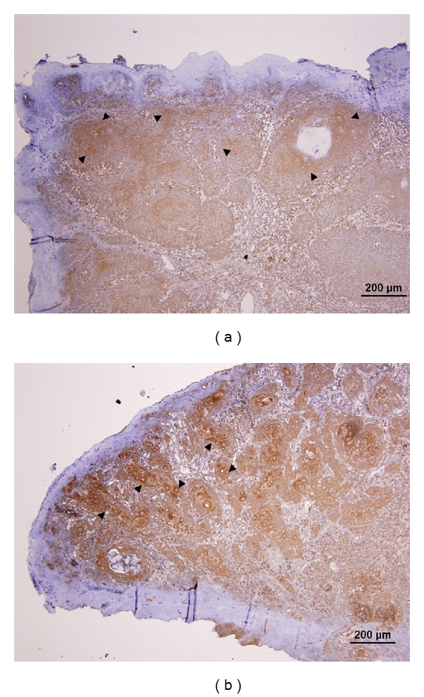
Immunohistochemical study on the expression of Akt1 and Akt2 from sections of an OSCC sample. Intense cytoplasmic staining of both Akt1 (arrowheads in (a)) and Akt2 (arrowheads in (b)) is observed in the tumor cells, while the overlying epithelium is principally negatively stained.