Fig. 1.

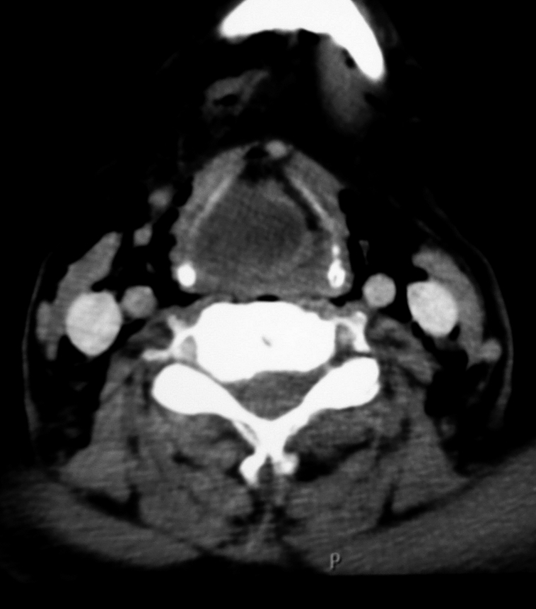
CT scan axial view demonstrating a supraglottic cystic hypodense mass with thickening of the wall. The mass causes an almost complete airway obstruction. It is confined to the larynx and a diagnosis of internal laryngopyocele was made.

CT scan axial view demonstrating a supraglottic cystic hypodense mass with thickening of the wall. The mass causes an almost complete airway obstruction. It is confined to the larynx and a diagnosis of internal laryngopyocele was made.