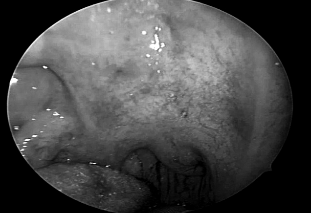
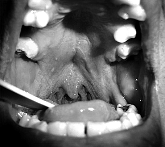
Fig. 2.


In, A. pre-operative view of a "excess" soft tissue structures is shown within the oropharynx upon oropharyngeal inspection during maximal mouth opening with the tongue relaxed in the mouth. Note the velar collapse at the level of posterior oro-pharyngeal wall; in B. a post-operative view is seen (followup: 14 months) showing retraction of the soft velo-uvulo-pharyngeal tissues with enlargement of the mesopharyngeal space, and complete visualization of the posterior pharyngeal wall.
