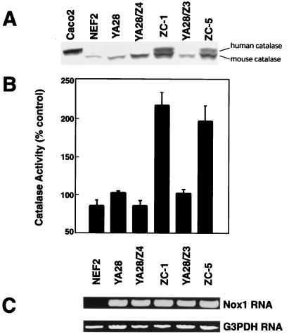
Figure 2.
Catalase expression and activity in cells transfected with Nox1 and h-catalase. (A) Catalase expression was determined in the indicated cell lines by Western blotting by using an antibody that recognizes both the human (upper band) and mouse (lower band) catalase. Caco-2 cells serve as a positive control for expression of h-catalase. (B) Catalytic activity (100% = 6.5 units/mg cell protein) was assayed in cell lysates. The mean and SE of 4–25 determinations is shown. (C) Reverse transcription–PCR demonstrates Nox1 expression. G3PDH (glyceraldehyde 3-phosphate dehydrogenase) serves as a loading control.

