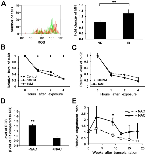
Figure 6.
Increased generation of ROS in the bystander cells and the effects of NAC. (A) An elevated level of ROS in bystander cells. Lin−Sca-1+ BM cells transplanted into IR or NR mice were collected 17 hours later and subjected to ROS measurement. Top panel: Representative flow chart. Bottom panel: Fold change of mean fluorescence intensity (MFI).**P < .01. n = 3. (B-C) Down-regulation of c-Kit expression at the protein and transcript levels by exposure to hydrogen peroxide. (B) c-Kit–enriched BM cells were exposed to different doses of hydrogen peroxide. Cells collected at different time points after exposure were subjected to c-Kit analysis by flow cytometry or RT-PCR. The relative expression of c-Kit at the protein level (B) and the transcript level (C) compared with nonexposed controls was plotted. (D) An elevated level of ROS reversed by administration of NAC in vitro. Lin−c-kit+ cells plated onto an IR (12 Gy) or NR stromal cell line (AFT024) layer treated with or without NAC were collected 40 hours later. The intracellular level of ROS was compared among groups. **P < .01. n = 3. (E) Improved reconstitution of bystander hematopoietic cells by administration of NAC in vivo. Homed Lin−c-kit+ cells in NR or IR recipients treated with or without NAC were sorted 17 hours after transplantation and retransplanted into lethally irradiated congenic recipients (3000 cells/mouse). Engraftment ratios of donor cells from IR recipients to that from NR recipients were calculated; n = 3. *P < .05. n = 4.

