Figure 1.
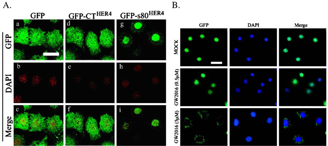
Nuclear localization of HER4/ErbB4 in mouse HC11 cells. a Localization of GFP fluorescence in HC11 cells stably expressing GFP, GFP-CTHER4 (the C-terminal residues 989–1308 of HER4/ErbB4, lacking the kinase domain), or GFP-s80HER4. b HC11 cells stably expressing GFP-s80HER4 were mock treated or treated for 24 h with 0.5 µM GW572016 (a dose that does not abolish s80HER4 tyrosine kinase activity) or 5 µM GW572016 (a dose sufficient to substantially reduce s80HER4 tyrosine kinase activity). The counter-stain with DAPI is shown as is the merge of the two images (GFP and DAPI). Bar=30 µm, for both (a) and (b).

