Figure 4.

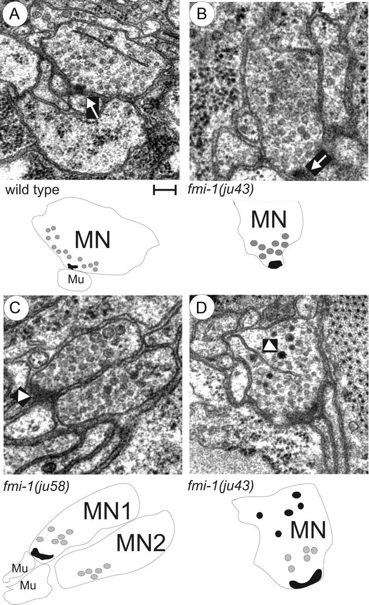
Ultrastructural analysis of fmi-1 synaptic defects. A, Wild-type animal NMJs formed as a single active zone (arrow) surrounded by evenly sized, clear synaptic vesicles. Below, a schematic is provided to illustrate the motorneuron (MN) and muscle (Mu). The active zone (black object) was surrounded by synaptic vesicles (gray circles), only some of the vesicles are illustrated for simplicity. B–D, The fmi-1 mutants did have some normal NMJs (B). However, many fmi-1 animals also exhibited abnormal junctions (C, D). C, In this junction, two neurons had vesicle-filled swellings at the point of contact with the muscle; however, only one had an active zone (arrowhead). This is illustrated below where motorneuron 1 (MN1) has an active zone, but motorneuron 2 (MN2) does not, despite both having an accumulation of synaptic vesicles. D, Electron-dense vesicles were present at ju43 NMJs (arrowhead), but not in wild-type animals (A). In the schematic, the electron-dense structures are illustrated as black circles. Scale bar, 100 nm.
