
Figure 6.
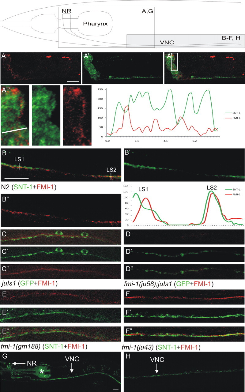
FMI-1 is concentrated at nonsynaptic regions of the nervous system. Using specific antisera, we visualized FMI-1 (A–F, red) relative to the synaptic vesicle protein SNT-1 (A, B, E, F, green) or juIs1 (SNB-1::GFP) (C, D, green). Above the panels is a schematic to orient the region being examined. A, B, In wild-type animals, FMI-1 localized to the nerve ring (NR; A) and ventral nerve cord (VNC; B). FMI-1 was predominantly adjacent to, but not overlapping with, SNT-1. A‴, Enlarged view of the boxed region in A″. FMI-1- and SNT-1-containing regions have little overlap. An intensity profile corresponding to the line in A‴ is on the right. B, In the ventral nerve cord, FMI-1 was adjacent to SNT-1 [line scan 1 (LS1)] but occasionally overlapped [e.g., line scan 2 (LS2)]. C, FMI-1 was adjacent to, but not within, GABAergic NMJs labeled by SNB-1::GFP. D, The ju58 allele lacked FMI-1 immunoreactivity, demonstrating the antisera specificity. Results were similar for tm306 and rh308 (data not shown). E, F, The missense mutations gm188 (E) and ju43 (F) did not grossly affect protein localization, although there is a slight increase in the overlap of the FMI-1 and SNT-1 (colocalization coefficient, 0.38). G, H, FMI-1::GFP accumulated in the NR and VNC in a pattern similar to the antisera staining. The pharyngeal staining (asterisk) is from the coinjection marker (Pmyo-2-mCherry). Scale bars, 10 μm.