
Figure 8.
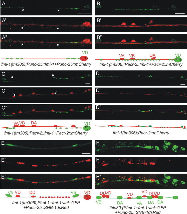
FMI-1 functions cell nonautonomously in the VD neurons. A, VD SNB-1::GFP puncta in fmi-1(tm306 + Punc-25::fmi-1 were enlarged and still had gaps (between arrowheads). A′, A″, RFP was used to mark cells with the transgene. Below is a diagram illustrating the VD neuron (red) expressing FMI-1 [F(+)], but long gaps were present lacking SNB-1GFP. B, When fmi-1 is specifically expressed in the cholinergic cells, SNB-1::GFP puncta were evenly sized and spaced puncta. B′, B″, RFPs, the transgenically positive neurons. C–C″, In some cases, the cholinergic (red) and GABAergic (green) axons appeared defasciculated. Here, even though the fmi-1 transgene is present in these cholinergic neurons, the GABAergic synaptic defects are not rescued. D–D″, Defects in SNB-1::GFP (green) were present when cholinergic axons (red) were properly fasciculated with the GABA neurons. E–E″, FMI-1ΔInt::GFP expressed under its endogenous promoter partially rescued the synaptic defects in GABAergic neurons (as visualized by SNB-1::RFP; E′). GFP is not observed in the GABAergic neurons. F–F″, We integrated the FMI-1ΔInt::GFP and still found no evidence of expression with the GABA marker (Punc-25::SNB-1::dsRED), confirming that FMI-1 expressed from non-GABAergic neurons is sufficient to rescue the SNB-1 accumulation defects. Scale bars, 10 μm.