
Figure 3.
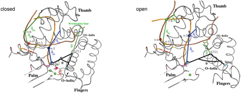
θ, δ, and β order parameters (see text and Supplemental Data for the definition), which describe the overall O-helix rotation, the DNA displacement, and the insertion of the preinsertion base into the preinsertion site, respectively, are shown in the closed and open conformations. The angle θ is defined as the angle (colored black) between Cα atoms of residues R629, G711, and N700 shown as cyan spheres. β (colored green) is the distance between Cα atom of G711 and the center of mass (C.O.M.) of the preinsertion base shown as green sticks. δic and δio measure the difference in the distance of a single phosphate atom from appropriate residues in the pol-I closed and open conformations, respectively. The residues contributing to δi of a given phosphate atom (n, n-3; n-3, n-4, n-6; shown as spheres) have the same color as that atom (see Table S1 for the list of residues). Brown and orange tubes represent template and primer strands, respectively. Preinsertion base and incoming nucleotide are shown as sticks and lines, respectively. Mg2+ ions and atoms defining the θ angle are shown as green and cyan spheres, respectively.