
Figure 1.
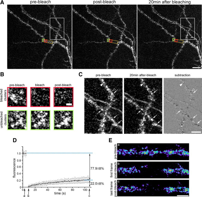
FRAP analysis reveals a stably clustered and a minor mobile fraction of CaV1.2-SEP in dendrites of hippocampal neurons. A, Representative image of a branched dendrite expressing CaV1.2-SEP before, immediately after, and 20 min after photobleaching (area indicated by yellow frame). Over 20 min the staining of CaV1.2-SEP clusters showed little changes outside and little recovery of fluorescence inside the bleached region. Scale bar, 10 μm. B, Enlargement of CaV1.2-SEP clusters in an unbleached (green frame) and a bleached (red frame) region of the dendrite in A at the three time points. Partial recovery of fluorescence 20 min after bleaching is not accompanied by a reappearance of the clusters. Scale bar, 1 μm. C, Enlargement of unbleached area in A (white frame) and corresponding subtraction image demonstrate that most CaV1.2-SEP clusters did not change (arrows), while a few clusters disappeared (filled arrowheads) or appeared (open arrowheads) during the 20 min observation time. Scale bar, 5 μm. D, Average normalized FRAP curve from experiments in which the fluorescence recovery was recorded for 99 s at 1.5 Hz (mean ± SD; N = 17 recordings from three experiments). On average, the CaV1.2-SEP fluorescence in the bleached dendritic segments recovered by 22.8 ± 8%. E, Representative frames (a–c) of a CaV1.2-SEP expressing dendrite from the FRAP experiment in C show that the recovering fraction of channels were not clustered. Scale bar, 5 μm.