Figure 2.
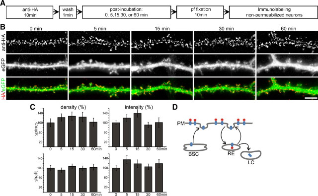
Surface-labeled CaV1.2-HA clusters in living hippocampal neurons are not internalized within 60 min. A, Experimental workflow. pf, Paraformaldehyde. B, CaV1.2-HA- and eGFP-cotransfected neurons were live cell labeled with anti-HA for 10 min and fixed 0, 5, 10, 15, 30, or 60 min after removing the antibody (antibody and subsequent incubation at 37°C). The secondary antibody was applied to unpermeabilized neurons to exclusively label channels remaining at the surface. Coexpressed soluble eGFP outlines the dendrite morphology. At all time points CaV1.2-HA clusters show a similar localization on dendritic shafts and spines. Scale bar, 5 μm. C, Quantification of the density (number of clusters per spine or per μm2 of dendritic shaft) and the fluorescence intensity (average gray value) of CaV1.2-HA clusters expressed as percentage of surface labeling at the 0 time point. Over 60 min no significant changes were observed (one-way ANOVA, p = 0.17–0.88). D, Schematic turnover pathways of membrane proteins. PM, Plasma membrane; BSC, biosynthetic compartment; RE, recycling endosomes; LC, lysosomal compartment. Bar graphs, mean ± SEM; N = 12–14 analyzed dendritic segments in three separate experiments.

