
Figure 7.
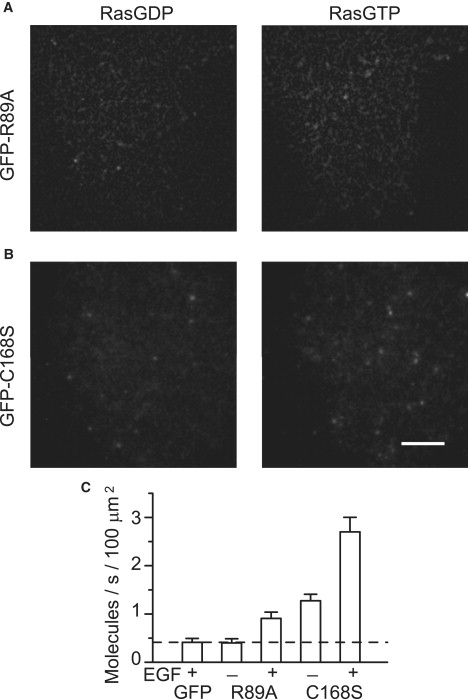
Single-molecule imaging of RAF mutants. (A) R89A and (B) C168S mutants of RAF were tagged with GFP and expressed in HeLa cells with Ras. The cells were observed by TIRF microscopy before (left) and after (right) stimulation with EGF to induce Ras activation (scale bar: 5 μm). (C) The averages of the molecules recruited to the basal cell membrane per second per 100 μm2 area of the membrane are shown with their standard error.