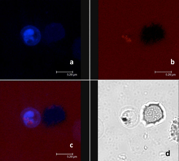
Figure 4.
Confocal microscopy analysis after double staining cells with nitidine and lysotracker®. a: staining with 120 μM nitidine, b: staining with 5 μM lysotracker®, c: merge, d: daylight. The repartition of nitidine was not similar to that of Lysotracker®, a stain that concentrates in the food vacuole of the parasite.

