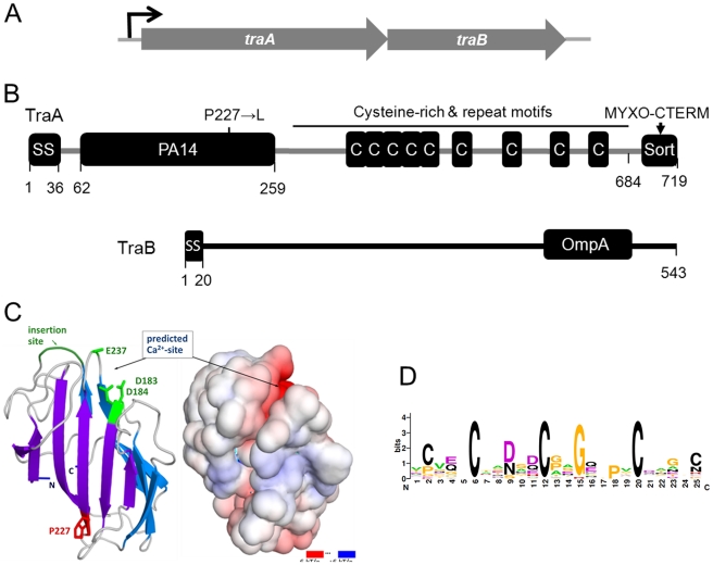
Figure 4. Genetic and modular structure of TraAB.
(A) Operon structure depicting genes that translationally overlap. (B) Domain and motif architecture and the DK396 amino acid substitution indicated. (C) Modeled three-dimensional structure and electrostatic surface potential of TraA PA14 domain. Features shown in green in the ribbon diagram (left) could serve to recognize glycans through potential side-chain coordination of a calcium ion by Asp183, Asp184, Glu237 (only Cα-Cβ shown), and the location of an insertion important for carbohydrate-binding specificity in FLO5 [24]. Graphics produced with PyMOL (Molecular Graphics System, Version 1.3, Schrödinger, LLC) and APBS Tools2 [61]. (D) consensus sequence LOGO [62] for Cys-repeats found in TraA and myxobacteria family members designated TIGR04201.

