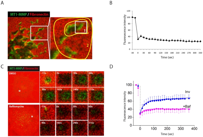
Figure 2. MT1-MMP transport to invadopodia via lysosomal secretion.
(A) MT1-MMP-phLuorin-expressing SCC61 cells cultured on Dylight 633-labeled fibronectin were subjected to FRAP- continuous photobleaching experiments. One half of the invadopodia area, indicated by the open box area in region 1, was the FRAP experimental area. Region 2 was a continuous photobleaching area. (B) Quantification of fluorescence recovery in the Figure 2A FRAP region is calculated. (C) Representative images of FRAP experiments at invadopodia without or with bafilomycin. (D) The recovery of FRAP signals are shown in the absence (blue circles) and in the presence (pink circles) of bafilomycin. Reconstructed time courses of fluorescence recovery in the absence (blue line) and in the presence (pink line)of bafilomycin at invadopodia are also shown. The reconstructed FRAP signals show a good agreement with experimental data.

