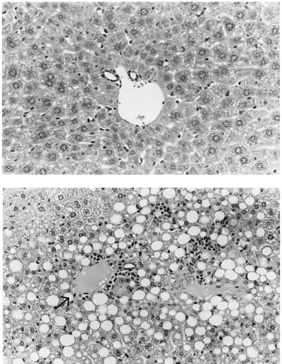
Figure 5.
Liver histology from 8-month-old MAT1A knockout mice fed a normal diet. (Upper) Wild type. The portal tract demonstrates a portal vein and two small duct structures. No portal or lobular inflammation or lobular fatty change is seen. Hematoxylin and eosin × 148. (Lower) MAT1A knockout mouse. The portal tract at the left of the field (arrow) shows mild portal lymphocytic infiltrates with an unremarkable bile duct. The hepatocytes adjacent to the portal tract show macrovesicular fatty change. In addition, focal areas of inflammation are also seen involving the liver cells that contain fat (steatohepatitis); the inflammatory component is chiefly lymphocytic. Hematoxylin and eosin × 148. Representative histologic changes are shown from three knockout mice.

