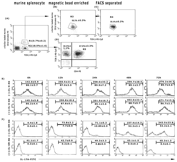
Figure 6. Kinetics of IL-17A production by iNKT cells of cured mice.
Sixty days LD infected animals were treated with GSPL as described in the legend of Figure 1. Animals were sacrificed 15 days after the last treatment. NKT cells from spleens of individual experimental BALB/c (A) mice were identified as α-GC/CD1d tetramer+TCRβ+ cells and after enrichment by magnetic cell sorting (B) were further purified by FACS sorting (C). The NKT− populations were identified as TCRβ+CD4+ cells (D). Isolated (E) iNKT cells or (F) iNKT depleted cell populations (R5, TCR-β+CD4+) were co-cultured with 1∶10 of autologous splenic adherent cells as described for Figure 4. Cells were stimulated with GSPL at 100 µg/mL, ± IL-6 Ab (E) or ± IL-6R Ab (F) for the time periods indicated and intracellular IL-17A was assessed using FACS analysis. Numbers above the horizontal bar indicates the mean fluorescence intensity and number below the bar indicates the percentage of IL-17 positive cells in the gated populations. Results show mean ± SD of three individual mice per group; paired two-tailed Student's t-test. Results of one from three independent experiments are shown.

