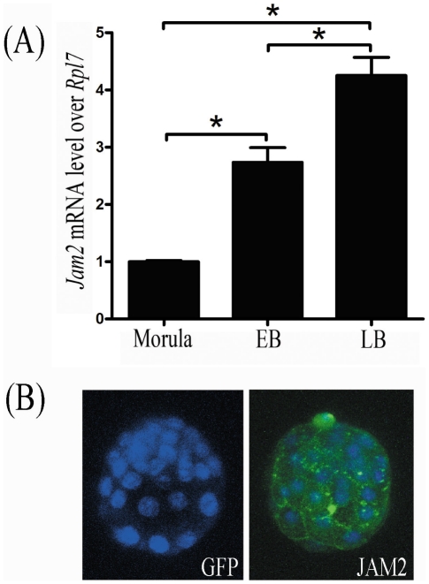
Figure 6. Jam2 expression in mouse embryos.
(A) Real time RT-PCR of Jam2 mRNA expression in mouse embryos at morula, early (EB) and late blastocyst (LB) stages, respectively. (B) The immunofluorescent analysis of JAM2 protein in mouse blastocysts. Anti-GFP antibody was used as negative control. Blastocyst nuclei were counter-stained with DAPI (Blue staining).

