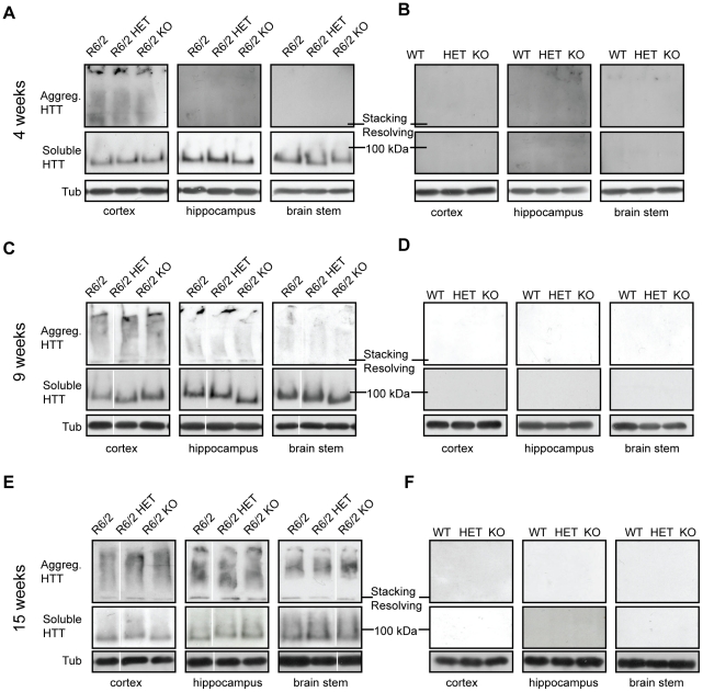
Figure 5. Levels of soluble mHTT in various brain regions at 4, 9 and 15 weeks of age.
Representative western blots from cortex, hippocampus and brain stem of (A–B) 4, (C–D) 9 and (E–F) 15 week old wild type (WT), Sirt2HET (HET), Sirt2KO (KO), R6/2, Sirt2HETxR6/2 (R6/2 HET) and Sirt2KOxR6/2 (R6/2 KO) mice probed with an anti-HTT antibody (S829) and tubulin (Tub) as loading control. Both soluble mHTT transprotein and aggregates retained in the stacking gel can only be detected in mice expressing the R6/2 transgene (A, C, E). All samples were run on the same gel. White lines indicate where lanes are not contiguous.

