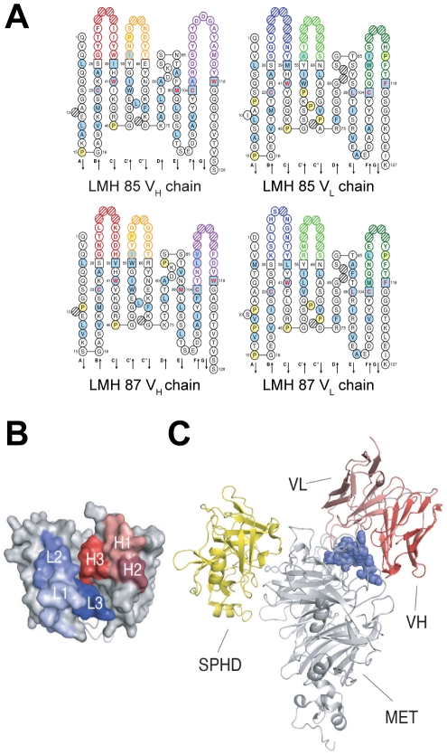
Figure 4. Docking of LMH 87 to c-MET.
(A) predicted amino acid sequences of LMH 85 and LMH 87 VH and VL domains. (B) 3D model of the VH and VL domains of LMH87 (modeled as a scFv). The complementarity determining regions (CDR) of the VH and VL domains are shown in red and blue respectively. Framework regions are shown in grey. (C) binding locations within MET567 (grey) of the serine-protease homology domain (SPHD) of HGF/SF (yellow) (PDB accession 1SHY) and the scFv LMH 87 (red). The location of the latter was obtained by docking the model of the LMH 87 scFv onto its epitope (blue spheres). The figure was drawn with PYMOL.

