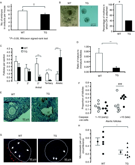
Figure 3.
Changes in the embryos of the TG female mice over-expressing VEGF165b. (A) Mice were culled at 0.5 dpc and embryos were collected from the oviducts. VEGF165b over-expression led to a reduced number of embryos in the oviducts. †P<0.05, Wilcoxon signed-rank test. (B) Images of embryos showing that those from TG females lack the corona radiata (CR) structure. #P<0.05 Fisher's exact test. (C) Ovaries from these mice were fixed, sectioned through and stained with H&E to count follicles. P<0.001 one-way ANOVA, *P<0.05, **P<0.01, ***P<0.001 Bonferroni post-hoc test. n=6 per group. (D) The number of tertiary follicles as a percentage of secondary follicles was determined. **P<0.01 Mann–Whitney t-test. (E) Ovaries were stained with cleaved caspase-3 antibody and the number of positive (apoptotic cells) was determined. (F) The proportion of follicles with between 1 and 10 and >10 positive cells were compared (###P<0.001, Fisher's exact test on follicle frequency, n=6 animals per group, 274 follicles WT, 234 TG) (G). Ovaries taken at 0.5 dpc were fixed and stained using isolectin B4 to detect endothelial cells, and counterstained with Hoechst. Images of follicles from WT and TG female ovaries stained with IB4 to show MVD. (H) Microvessels were counted and expressed as microvessel covering area per unit perimeter of the follicle. *P<0.05, Mann–Whitney t-test.

