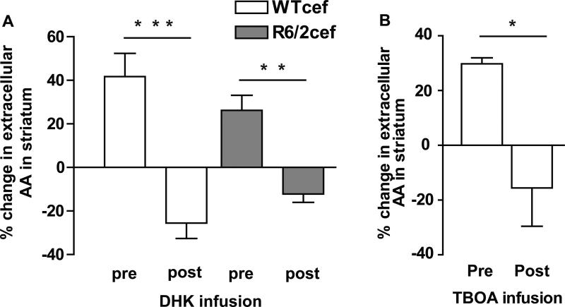
Figure 4.
Intrastriatal infusion of either DHK or TBOA blocked evoked levels of AA in striatum. X-axis labels for A and B represent data collected with the cannula placed in striatum but before infusion (pre), and data collected following infusion (post) in the same animals. (A)There was a main effect of infusion [F(1,16) = 49.6, p < 0.001]. No effect of genotype emerged [F(1,16) = 0.02, p > 0.05]. Note the similarity between the WTcef and R6/2cef data here compared to Fig. 2. A Tukey test indicated that DHK infusion diminished striatal AA in WTcef (***p < 0.001) and in R6/2cef mice (**p < 0.01) compared to the pre-DHK condition. (B) TBOA also reduced evoked release of AA in WTcef striatum (t-test, *p< 0.05). n = 8 for WTcef and n = 6 for R6/2cef in DHK experiments, and n = 3 for WTcef TBOA experiments.

