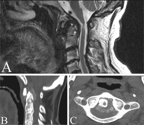
Fig. 2.
Preoperative neuroimaging studies. a, b Sagittal T2 MRI image and sagittal CT scan showing bulbo-medullary compression by rheumatoid pannus and the associated area of myelopathy. Cervicomedullary angle (CMA) measured on sagittal T2 MRI image was 137°. c Axial CT scan reveals the odontoid process with its backward rheumatoid pannus

