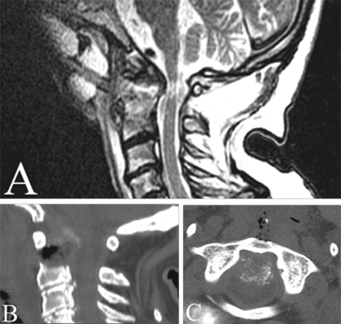
Fig. 3.
Postoperative neuroimaging studies. a Sagittal T2 MRI image showing adequate spinal cord decompression with an improved CMA (151°) and realignment of the medulla. After the decompression, the area of myelopathy was better visualized. b, c Sagittal and axial CT scans showed the odontoidectomy emphasizing the anterior C1 arch integrity

