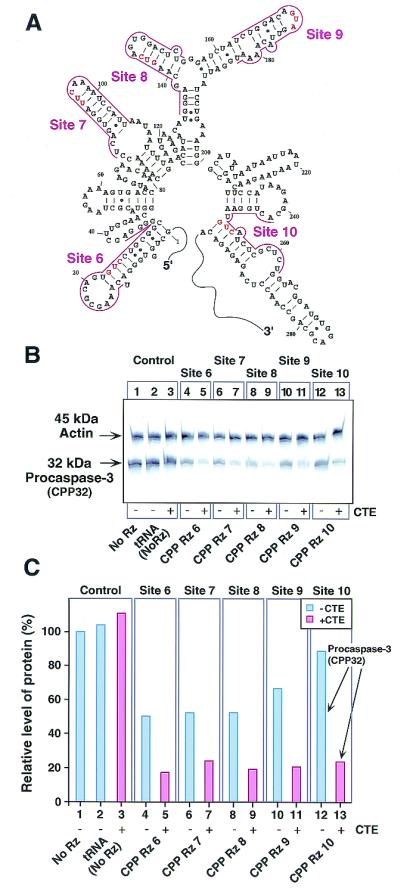
Figure 4.

Inhibition of procaspase-3 (CPP32) gene expression by CTE-Rz. (A) The secondary structure predicted by MulFold (19) of the 5′ region of procaspase-3 mRNA targeted by Rz. (B) Detection of procaspase-3 and actin proteins by Western blotting (10). Mouse NIH 3T3 cells were transfected with the indicated Rz constructs. (C) The results in B presented as a histogram. FITC-labeled antibodies against rabbit IgG were used as the secondary antibody, and the band intensities for actin and procaspase-3 were quantitated. Procaspase-3 protein levels were normalized to actin protein levels. The normalized level of protein recorded when cells were untransfected with the Rz-expressing vector was taken as 100% (lane 1).
