Fig. 4.

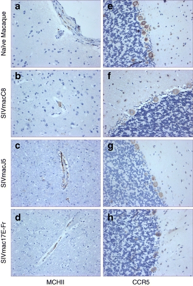
Representative images showing immunohistochemical staining results for a–d MHCII (×20) and e–h CCR5 (×20); a–d frontal lobe and e–h cerebellum of either SIV naive or SIV-infected brain samples

Representative images showing immunohistochemical staining results for a–d MHCII (×20) and e–h CCR5 (×20); a–d frontal lobe and e–h cerebellum of either SIV naive or SIV-infected brain samples