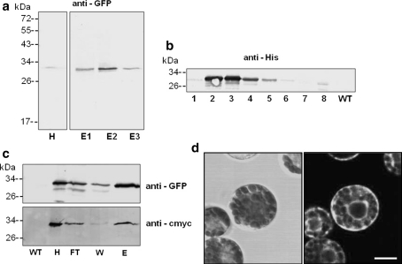
Fig. 5.
GFP-expression in transformed tobacco plants. a Leaves of a transformed tobacco plant (Agro-injection) were homogenised after 5 days, purified via Ni–NTA agarose and detected by an anti-GFP antibody (H: 40 μg, E1–E3: 4 μg protein per lane). b Leaf homogenates of various stable transformed plants of the T1 generation cultivated on kanamycin-containing media (numbers indicate individual plants) and wild type (WT) were prepared and analysed by immunodetection using an anti-His antibody (15 μg protein per lane). c Detection of GFP in leaves harvested from tobacco plants of the T3 generation. GFP was purified from leaf homogenates by affinity chromatography (Ni–NTA IDA) and detected by an anti-GFP as well as an anti-cmyc antibody directed against the C-terminal cmyc-tag (27, 13, 6 and 1 μg protein per lane for fractions H, FT, W and E, respectively). d Mesophyll protoplasts prepared from leaves of transformed tobacco plant were observed by confocal laser scanning microscopy. Bright field image on the right and corresponding fluorescence image (left) Bar 10 μm. All plants were transformed with pBINPLUSpIMPACT-GFPER. WT wild type homogenate, H homogenate, FT flow through of column, W wash fraction, E eluate

