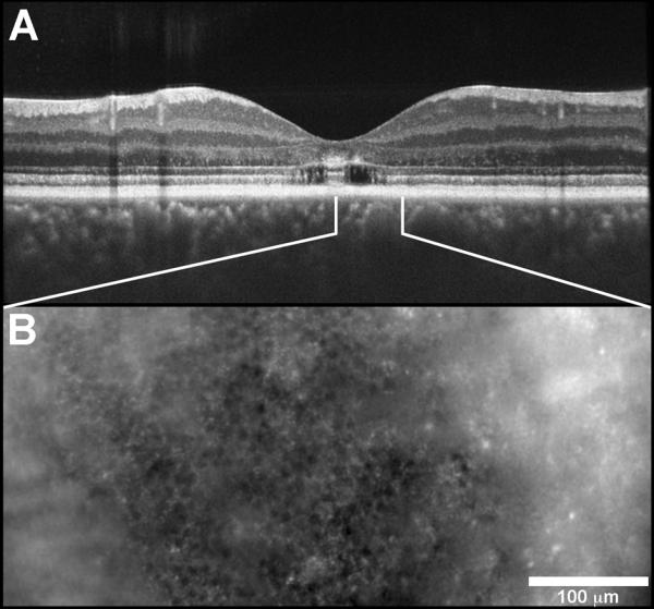
Fig. XX.2.

Image from patient with a bulls’s eye maculopathy. (A) SD-OCT scan shows that retinal layers are preserved in a small island in the central macula with loss of the IS/OS layer and the ELM in the perifoveal region. The white lines indicate the area imaged with AO in (B).
