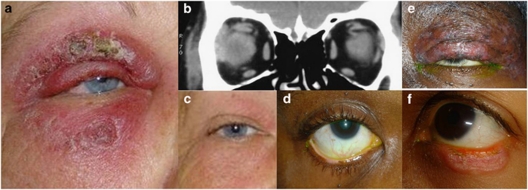
Figure 1.
(Patient1) (a) Violaceous swelling with eczematous changes at presentation; (b) Coronal orbital CT images demonstrate right lacrimal gland, superior and lateral recti enlargement. (c) Facial swelling almost completely resolved without scarring; (Patient 2) (d) Localised lower lid margin depigmentation, preserved lashes, no erythema; (Patient 3) (e) Diffusely bulky left upper eyelid with areas of depigmentation and atrophy; lateral lid margin destruction and madarosis. (Patient 4) (f) Lower lid at presentation shows erythematous, scaly, discoid lesion with madarosis.

