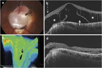
Figure 2.
Patient 3, left eye. (a) Fundus photograph of the optic disc on presentation, demonstrating an inferior coloboma (black arrow). A retinal break at the location of the arrow is difficult to visualise clinically. (b) OCT of the macula on presentation, demonstrating retinoschisis (asterisk) with subretinal fluid (white arrow) and retinal tissue extending into the optic disc cavity (broken arrow). (c) 3D OCT reconstruction of the optic disc demonstrating a retinal break in the cup of the disc (black arrow). (d) OCT of the macula 9 months postoperatively demonstrating marked reduction of the macular schisis and subretinal fluid.

