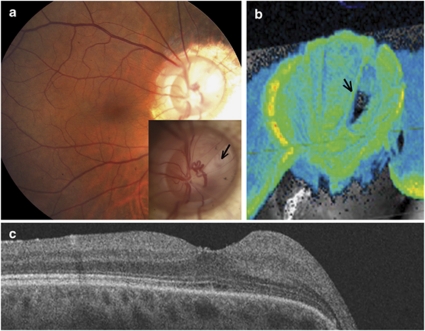
Figure 4.
Patient 5, right eye. (a) Fundus photograph of the macula 10 years postoperatively, demonstrating resolution of the retinal detachment. Inset, optic disc photograph, with a break in the thin overlying retinal tissue (black arrow). (b) OCT 3D reconstruction of the optic nerve head, showing a deeply excavated defect and an oval retinal break in the retina within the morning glory defect (black arrow). (c) OCT imaging demonstrating a completely attached retina after vitrectomy with separation of the posterior hyaloid, membrane removal, gas tamponade and laser photocoagulation.

