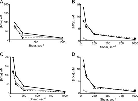
FIGURE 6.
Models of fIIa and fXa generation. fIIa and fXa generation under steady-state conditions were modeled using the bulk substrate diffusion model (Equation 12; gray line) and membrane-localized model (Equation 13; black line). Experimental data are shown with a dotted line. A, isolated bovine prothrombinase (23); B, isolated extrinsic tenase; C, coincident activation of hfII; D, coincident activation of hfX.

