Fig. 3.
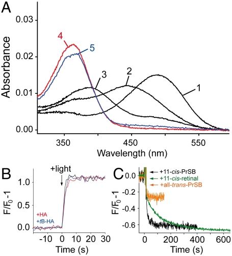
Characterization of mutant K296G. (A) UV/visible spectrum of K296G opsin regenerated with 11-cis-PrSB in the dark (1), 30 s (2) and 160 min (3) after illumination of the sample. Spectra (4) and (5) were recorded 30 s after illumination of samples containing 25 mM hydroxylamine or tB-HA, respectively. (B) Light-induced retinal release from K296G monitored by fluorescence spectroscopy in the absence (black trace), and in the presence of 25 mM hydroxylamine (red trace) or tB-HA (blue trace), respectively. (C) Fluorescence change induced by addition of 1 μM 11-cis-PrSB (black trace) or all-trans-PrSB (orange trace) to 0.5 μM K296G opsin. Addition of 1 μM 11-cis-retinal to control opsin (green trace). All measurements were performed at pH 6.0, 10 °C in 0.03% DDM.

