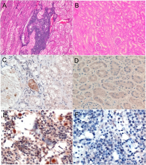
Fig. 4.
(A and B) Histological section of kidneys stained by H&E from a stray cat with FmoPV detected in urine and a normal cat, showing aggregates of inflammatory cells in the interstitium and renal tubular degeneration in the infected cat. (C and D) Immunohistochemical staining of kidney sections of stray cat with FmoPV detected in urine using guinea pig serum positive for anti-FmoPV N protein antibody and preimmune guinea pig serum, showing positive renal tubular cells. (E and F) Immunohistochemical staining of lymph node sections of stray cat positive for FmoPV using guinea pig serum positive for anti-FmoPV N protein antibody and preimmune guinea pig serum, showing positive mononuclear cells.

