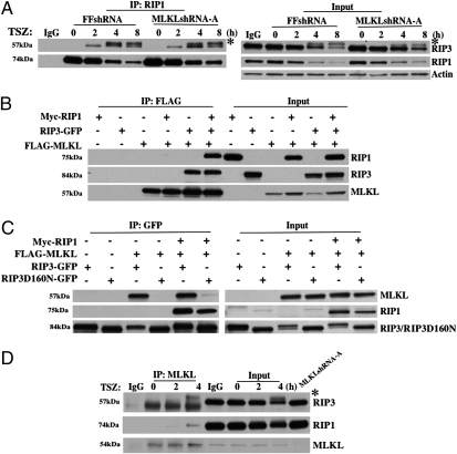
Fig. 2.
MLKL interacts with RIP3. (A) HT-29 clones FFshRNA and MLKLshRNA-A were treated with TSZ for the indicated times; cell lysates were immunoprecipitated with anti-RIP1 antibody (IP:RIP1) and analyzed by immunoblot with anti-RIP3, anti-RIP1, or anti-Actin antibodies. Input, 1% of extract before immunoprecipitation (control). * indicates phosphorylated RIP3. (B) HEK293 cells were transfected with Myc-RIP1, RIP3-GFP, or FLAG-MLKL as indicated. After 24 h, cell lysates were immunoprecipitated with anti-FLAG antibody (IP:FLAG) and analyzed by immunoblot with anti-Myc, anti-GFP, or anti-FLAG antibodies. Input, 3% of extract before immunoprecipitation (control). (C) HEK293 cells were transfected with Myc-RIP1, FLAG-MLKL, RIP3-GFP, or RIP3D160N-GFP as indicated. After 24 h, cell lysates were immunoprecipitated with anti-GFP antibody (IP:GFP) and analyzed by immunoblot with anti-FLAG, anti-Myc, or anti-GFP antibodies. Input, 3% of extract before immunoprecipitation (control). (D) HT-29 cell lysates were immunoprecipitated with anti-MLKL antibody (IP:MLKL) and analyzed by immunoblot with anti-RIP3, anti-RIP1, and anti-MLKL antibodies. Input, 1.5% of extract before immunoprecipitation (control). * indicates phosphorylated RIP3.

