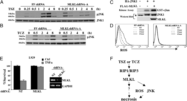
Fig. 4.
Loss of MLKL affects JNK phosphorylation. (A) HT-29 clones FFshRNA and MLKLshRNA-A were treated with TSZ for the indicated times; cell lysates were analyzed by immunoblot with anti-pJNK or anti-JNK1 antibodies. (B) HT-29 clones FFshRNA and MLKLshRNA-A were treated with TCZ for the indicated times; cell lysates were analyzed by immunoblot with anti-pJNK or anti-JNK1 antibodies. (C) HEK293 cells were transfected with either FLAG-vector, or FLAG-MLKL, and/or HA-JNK1 as indicated. After 24 h, cell lysates were immunoprecipitated with anti-HA antibody and used for in vitro kinase assay using GST-cJun (1–79) as substrate. Lysates were analyzed by immunoblot with anti-FLAG and anti-HA antibodies. * indicates nonspecific band. (D) HT-29 clones FFshRNA and MLKLshRNA-A were treated with TSZ for the indicated times, stained with DCFDA, and analyzed by flow cytometry. Gray line (NT) represents background ROS in untreated samples. (E) L929 cells were infected with lentivirus, nontargeting (NT) or MLKL shRNA for 24 h followed by selection with puromycin for 48 h. Cells were then untreated or treated with TNFα (30 ng/mL) for 8 h. Cell survival was determined by MTT assay. (Right) RT-PCR of L929 cells infected with NT or MLKL shRNA with MLKL or GAPDH primers. (F) Diagram of the role of MLKL in RIP1/RIP3-dependent necrosis.

